The European Network for a cure of ALS (ENCALS) held its 11th Annual meeting in Sheffield from 31 May to the 2 June. The weekend was full of glorious British sunshine and more than 200 international scientists and clinicians were also able to enjoy a range of incredibly interesting talks about the latest developments in MND research.
A particular talk caught my attention on the first day by Dr Johannes Brettschneider from the University of Ulm in Germany. Dr Brettschneider explained how his research had shown the stages and spread of the protein TDP-43 in ALS (the commonest form of MND).
Dr Brian Dickie, Director of Research Development, said: “The key to defeating MND lies in fostering strong collaborations between neurologists, healthcare professionals, research scientists, early career investigators and students in the field of MND and the 11th Annual ENCALS meeting in Sheffield provided that opportunity. The MND Association was proud to support this event.”
‘Special’ staining
At the end of an afternoon of talks on the MND- causing genes C9orf72, FUS and SOD1, Dr Brettschneider engrossed over 200 delegates with his talk on the TDP-43 protein and how it spreads in ALS.
Although TDP-43 genetic mistakes are a rare cause of MND, scientists are especially interested in the TDP-43 protein because in the vast majority of cases of MND (irrespective of whether it was caused by an inherited genetic mistake), TDP-43 protein forms pathological clumps inside motor neurons.
The study (which is a collaboration between Dr. John Trojanowski and Dr. Virginia Lee from the Penn University Center of Neurodegenerative Disease Research in Philadelphia, America and the group of Dr. Heiko Braak in Ulm) used a technique known as ‘immunohistochemistry’. This technique involves taking tissue samples of the brain and spinal cord from people who have died from ALS. The researchers would then make extremely thin slices of the tissue, which could then be stained using a ‘special stain’ and viewed under a microscope.
The stain used by Dr Brettschneider only ‘stained’ the TDP-43 protein in the samples, meaning that he could see the amount of TDP-43 in different areas of the brain and spinal cord.
Using the clinical information and TDP-43 staining this would allow Dr Brettschneider to stage the disease.

Axonal ‘telephone wires’ do more than just talking
Dr Brettschneider showed that TDP-43 increased in different areas of the brain and spinal cord during different stages of the disease. Amazingly, he also showed how ALS (characterized by clumps of TDP-43) spreads from one are of the body to another.
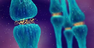
A motor neurone consists of three parts; the cell body, axon and nerve ending. The cell body contains the nucleus, or the control centre of the cell. When a message travels from the brain the cell body sends the message down the axon. Like telephone wires, the axon carries the message to the muscle, where the nerve endings cause the muscle to move.
However, in ALS it seems that these ‘telephone wires’ do more than just carry a message. The protein TDP-43 forms ‘clumps’ in the motor neurones and it seems that these clumps use the axon to travel from one motor neurone to the next (possibly explaining why someone get’s weakness in their arm and then their hand).
Another key finding was that TDP-43 clumps develop in the front part of the brain (prefrontal cortex), which is responsible for personality and may explain the development of cognitive symptoms.
Dr Brettschneider explained the importance of this research “While spreading of disease-related proteins has been described for other neurodegenerative diseases like Alzheimer’s disease or Parkinson’s disease, this had not been previously shown in ALS. Now, we can show evidence that supports a spreading of the major disease protein TDP-43 in ALS across specific regions of the brain and spinal cord with ongoing disease.
If these findings can be confirmed (for example in cell culture or mouse model studies) then this could lead to the design of new treatments specifically aiming to impair the spread of TDP-43 protein clumps.

Furthermore, we believe that our findings offer a better understanding of disease progression in ALS. Our data implies that TDP-43 spreads throughout the prefrontal cortex with ongoing disease, thereby lending support to the idea that all ALS patients could eventually develop “frontal type” cognitive deficits.”
The future
Dr Brettschneider commented why this research is important to people living with MND explaining that “If these stages can be reproduced in patients with ALS they could offer a new way to assess disease progression and response to new treatments. We hope that our study provides the essential groundwork for strategies designed to prevent pTDP-43 spread.”
This research is only the beginning and more work is needed, Dr Brettschneider also explained what he hoped to do next with these exciting results. “There were restrictions in time and availability of the tissue samples during this study, so we were unable to determine how and where exactly ALS begins in the very early stage of the disease. Therefore, an important next step in our work would be to analyze very early cases with ALS to look at TDP -43 spread as this offers the most promising window for therapeutic intervention.”
Reference
Brettschneider J, Del Tredici K, Toledo JB, Robinson JL, Irwin DJ, Grossman M, Suh E, Van Deerlin VM, Wood EM, Baek Y, Kwong L, Lee EB, Elman L, McCluskey L, Fang L, Feldengut S, Ludolph AC, Lee VM, Braak H, Trojanowski JQ. Stages of pTDP-43 pathology in amyotrophic lateral sclerosis. Ann Neurol. 2013 May 20. doi: 10.1002/ana.23937. [Epub ahead of print]


Very nice read out! Thank you for posting!
Please god let this work x
Sounds very interesting progress and any progress is good. Unfortunately my husband died I. November due to MND.
Keep up this wonderful work!