 On 4 November, we welcomed two of our funded researchers to our offices in Northampton. Ruxandra Mutihac and Matt Gabel gave us a ‘taste’ of what’s to come for this year’s 25th International Symposium on ALS/MND, by speaking to us about their research.
The symposium is the World’s largest MND-specific research conference and is now only two weeks away! Find out more about the symposium here.
The abstracts (or research summaries), of all the research being presented during the symposium, are now available to view online via our website. On the 5 December we will also be reporting LIVE from the symposium in Brussels on this blog, so make sure you subscribe in order to get the reports delivered straight to your email inbox!
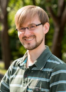
Matt Gabel
Association-funded PhD student, Matt Gabel, is attending his first symposium and will be presenting a poster about his work to over 800 researchers. Having recently only started his PhD this year, Matt is looking forward to presenting his initial work on applying maths to Magnetic Resonance Imaging (MRI) data. Matt is specifically looking at diffusion-weighted imaging, which he explains below:
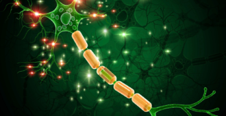
In MRI, the brain is divided into voxels or cubes – a voxel is the 3D equivalent of a pixel.
Diffusion-Weighted Imaging looks at the diffusion of the water molecules in each voxel of the brain.
Water in a free environment, such as corticospinal fluid, can diffuse easily in all directions. In biological tissue there are barriers, such as cell walls and nerve fibres, which reduce the ability of water to diffuse.
In grey matter within the brain, the biological barriers are spread out evenly, so the water molecules can diffuse evenly in all directions (we call this isotropic diffusion). White matter is made up of long thin fibres, and the fibres restrict diffusion so that it is mostly in one direction (which we call anisotropic diffusion).
We can exploit these differences in diffusion properties to reconstruct the paths of the white matter nerve fibre bundles, in a process called tractography to find out more about what happens in the brains of people with MND.
It’s amazing because I’ve done something theoretical and got something real out of it!
Ruxandra Mutihac
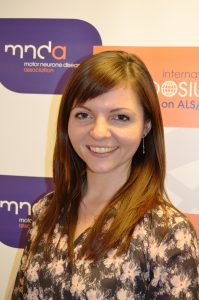
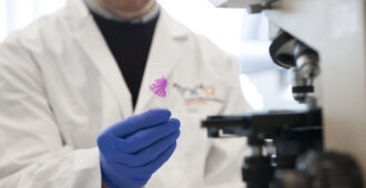
Dr Ruxandra Mutihac attended last year’s symposium in Milan, Italy, where she presented a poster of her work. This year, she has the daunting task of presenting her work as an oral communication on day two of the symposium! Ruxandra is using induced pluripotent stem cell technology to further our understanding of MND. This process of going back in time and creating stem cells is not as easy as it sounds, as she tells us below:
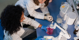
In our lab, we generate motor neurons from skin biopsies of people living with MND. The process involves two main stages: (1) the reversal of skin cells to pluripotent stem cells and (2) the generation of motor neurons from pluripotent stem cells.
To better understand this concept, we need to consider the fact that all cells in our body have originated from a stem cell. The stem cells are capable of becoming any type of cell with a defined function (heart cells, liver cells, neurons, etc). As the stem cell divides, the resulting cells become specialized to various functions according to the environmental cues.
For a long time, this process was believed to be unidirectional, but owing to revolutionary work in stem cell research we now know that the process can be reversed and we have the tools to reverse it. Therefore, we can take a specialized cell, such as a skin cell, and turn its memory back to a stem cell.
Once the stem cells are generated, we provide them with a specific combination of cues that spinal motor neurons would receive during development. At the end of the entire process, which can take between 6 to 8 months, we will have a dish full of motor neurons that carry the patient’s genetic background and that provides us with unlimited opportunities to study the disease directly in the cell type affected by MND.
Brussels
We will be reporting LIVE from Ruxandra’s presentation during the symposium on 6 December, and we will be aiming to cover as much as the symposium as possible! From biomarkers to genetics we will bring you the latest news from the symposium via this blog.


Hello MNDA team, I note that the first four posters in the “Therapeutic Strategies” session are all related to gene therapy. I’ve not really come across gene therapy as a therapeutic methodology for MND before not least, I assume, because for the majority of us it’s not really clear that a single gene is causing the problem.
The first poster in the list for this session is entitled “P264 GENE THERAPY FOR SPORADIC ALS USING AN INTRAVENOUS INJECTION OF AAV VECTOR ” and at first sight sounds incredibly exciting. Would it be possible for you to give us a summary of these four posters together with your objective take on whether they really are as exciting as they sound?
I’ll look forward to your summaries of the symposium in general with emphasis on anything that’s particularly exciting!
Cheers, Colin Fenwick
Hi Colin,
Thank you for your comment. I’m glad that you found the posters that are being presented in the ‘Therapeutic Strategies’ session of interest. I have some free time during Saturday’s poster session to write a report on this, and I will, of course, report on these four posters in particular and try and speak to the presenters for you! 🙂
Best wishes,
Sam
on behalf of the MND Association’s Research development team
hmmmm… Now a paper from a Chinese group also proposing gene therapy to correct ADAR2 disfunction http://www.karger.com/Article/FullText/368927 “Exploration of the Pathogenesis of Amyotrophic Lateral Sclerosis from the Perspective of Motor Neuron TDP-43 Protein Expression and ADAR2 Activity”. Something of a trend developing?
It would be good if that “dish full of neurons” could be transplanted into the brain of a MND patient.. it’s too late for my beautiful son but I am terrified for his children and maybe their offspring. Why have no tests been done on the children to identify the faulty gene I wonder.
Dear Christine,
While pre-symptomatic testing for close relatives of someone with MND is possible, it is often only done in cases where a genetic mutation was identified in that person. Because MND is likely to be caused by a collection of different factors (and one of these is likely to involve genetic mistakes), finding a genetic mistake related to MND in that relative (ie your son’s children) does not necessarily mean that they would go on to develop the disease. This is why pre-symptomatic genetic testing in MND is something that should be thought about very carefully as it can have an immense impact on your family’s lives. I very much recommend that you read through our information sheet B2: genetic testing. You can also contact us directly for more information on this issue at research@mndassociation.org or mndconnect@mndassociation.org for a more general support.
Best Wishes,
Martina