Brain banks are a vital resource in MND research. The MRC London Neurodegenerative Diseases Brain Bank was established in 1989. It is part of King’s College London and King’s College Hospital, and is part-funded by the Medical Research Council (MRC).

After 18 months of planning, the bank has recently relocated into a bright terracotta building, fit with state-of-the-art equipment and plenty of space to teach in.
To celebrate the move, my research team colleague Martina and I attended their open day. We heard some interesting talks then got to meet the team, tour the labs, and even see a brain dissection! Here’s what we found out…
What is a brain bank?
A brain bank collects and stores tissue of the central nervous system (the brain and spinal cord) for use in research. It distributes tissue samples to the research community to improve our understanding about neurodegenerative diseases like MND, Alzheimer’s and Parkinson’s.
Healthy tissue (from people with no such diseases) is also stored – it’s just as important as tissue affected by disease because researchers always need to compare their results with healthy tissue.
How is tissue collected and stored at the brain bank?
Tissue is collected as soon as possible after death. Because of this, someone who wants to donate their tissue must make everyone aware of their wishes beforehand. They will also need to contact a brain bank and give their consent. When that person dies, the brain bank is contacted as soon as possible and arranges for specialist courier services to collect the tissue, ideally within 48 hours.
At this brain bank, half of the tissue is sent for analysis and the other half is frozen and stored at -80ᵒC – much colder than a freezer at home. The team at the bank analyse the tissue and add a pathological diagnosis to their database (these steps are described below). If they wish, the family is also informed of their findings.
How is the tissue analysed at the brain bank?
Firstly, the tissue is weighed. Often in diseases that affect the brain, the brain weighs less than average (the average weight is around 1.3 kilograms) due to degeneration of the nerve cells.
The next step is to look at the tissue. We were shown a brain that was affected by Alzheimer’s disease with possible Lewy Body dementia – the researcher could confirm this simply by looking at the shape and size of the parts of the brain that would be affected, without cutting it whatsoever.
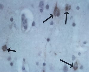
The brain was then carefully sliced, and 16 key parts from the slices were selected for further examination. These parts would then be ‘stained’ with special chemicals to help confirm if the tissue is affected by a disease – for example, a stain that picks up TDP-43 protein clumps might be used to confirm MND (these clumps are found in almost all cases). As the spinal cord is also affected in MND it would also be looked at, if possible.
This work is very important so that an accurate description can be provided for future research.
How is tissue used in research?
The findings from the tissue examination are recorded onto a database, which researchers all over the world can access, with permission. They have to apply to a special committee and explain in detail which samples they want to use and why they want to use them in their research.
If the application is approved, the appropriate samples are selected, packaged and sent off. The researchers report back to the tissue bank on their results.
How much tissue does a research project need?
One brain or spinal cord is used for many different research projects. Often, researchers use less than one gram of tissue for analysis (this means that one brain can provide more than 1000 samples!) We found out that this bank collects about 100-120 brains a year (around 10 from people with MND). In 2016, 11 out of 54 projects that used the bank used MND tissue – approximately 1,200 MND samples were used.
How useful is tissue in MND research?
Well characterised tissue is one of the most important resources in MND research. Just this year, genetic research using tissue from this bank helped to confirm the NEK1 gene as a cause of MND. Tissue allows researchers to observe what happens in the neurones, to help us understand how MND develops and find targets for drug development. Researchers can also use the tissue to see the effects of therapies in clinical trials, and to search for biomarkers of disease.
A collaborative network of brain banks
The key to defeating MND lies in collaboration. This bank is part of a larger initiative, led by the MRC, which aims to establish a coordinated national network of UK brain tissue resources for research. It is also part of the Brains for Dementia Research network, which aims to encourage and facilitate both tissue donation and accessibility and use by researchers.
Want to find out more?
More information about donating your tissue for MND research is available in our research information sheet. It explains more about the process and what to consider if you are thinking of donating your tissue.
We would like to thank the team at King’s for such an interesting and informative day.


This sounds amazing, I know that my son, who passed away in February this year, would have donated and I would have been very interested in the findings as I am convinced that Adam’s MND was triggered by high protein, high glutamate health supplements. Unfortunately this is the first time I have heard of the brain bank. We were never asked about organ donation of any sort. We gave blood and DNA early on but this was attached to a clinical trial for Dexpramipexole. It was also anonymised, which I thought strange, as we are told that many factors can be involved in neuron degeneration and so it seems that a full patient screen would be helpful for research purposes.