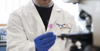
Researchers from University College London led by Dr Pietro Fratta and Dr John Thornton found that muscle imaging can help distinguish Amyotrophic Lateral Sclerosis (ALS) from Kennedy’s Disease based on the way specific muscle groups deteriorate in each condition. The method can also help assess the severity of the disease.
ALS is a rapidly progressing condition which affects both upper and lower motor neurones, leading to inability to move limbs and failure of breathing muscles at the later stages of the disease. The complex cause of this condition is not yet fully understood but is thought to be a combination of genetic and environmental factors. Kennedy’s Disease on the other hand is much slower in progression and severity and is primarily caused by a gene mutation.
Although separate motor neurone diseases with different causes, ALS and Kennedy’s Disease may share some symptom similarities. Having a measure that would provide a straightforward distinction between the two would shorten the time to diagnose the correct condition and potentially offer a way to track disease progression in clinic as well as treatment trials.
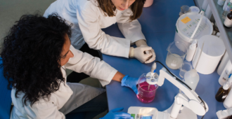
In the study, 21 people with ALS and 21 with Kennedy’s Disease were recruited alongside of 16 control participants to have their thighs, calves and head-neck regions scanned using muscle Magnetic Resonance Imaging (MRI). Paper-based functional rating scales, which are currently utilised in clinical trials, were used to measure the functional ability of people with ALS and Kennedy’s Disease, and matched to their unique muscle pattern.
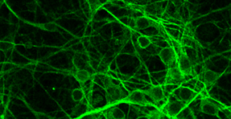
The main focus was on the proportion of muscle to fat tissue as well as accumulation of fluid within the muscle, with small sections of each muscle group blindly and independently rated for fat infiltration by clinicians and a computer software.
Although muscles were affected in both patient groups, the specific pattern varied significantly. In people with ALS, there were signs of built up fluid within the lower limb muscles, representing denervation of the muscles and accumulation of fluid from the diseased motor neurones. Meanwhile, in people with Kennedy’s disease, there was a significant fat infiltration in their bulbar and lower limb muscles, which are evident on the image below.

Although other tests exist that distinguish ALS from Kennedy’s disease, especially a blood test to reveal presence of a genetic mistake linked with Kennedy’s Disease, some people with this disease are still initially diagnosed with ALS. Including muscle MRI in the initial workup might help differentiate these conditions sooner.
“We were very surprised to see that some specific muscles seem to be spared in disease. Understanding why this happens could allow to identify protective factors from disease”. Dr Pietro Fratta
An even greater benefit of this technique might be its ability to track the progression of the disease, which was shown by relating the specific muscle patterns to the established functional rating scales.
With the current lack of biomarkers, muscle MRI has the potential to track changes in the muscles over time, tracking whether or how fast the disease progresses. This would enable shorter trials to be undertaken, reducing costs and allowing more therapies to be tested. As this was a ‘snapshot’ study however, further longitudinal studies need to be conducted to establish this method as an effective and sensitive biomarker of ALS and Kennedy’s Disease.
You can read the full paper here:
https://n.neurology.org/content/neurology/early/2019/08/06/WNL.0000000000008009.full.pdf

Nice article
Hi Marie,
Thanks for taking the time to read our blog post and leave a supportive comment.
Kind regards,
Research Information Team