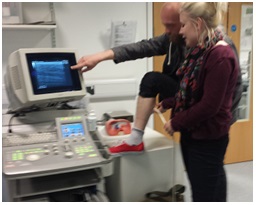
Kate Bibbings is a PhD student funded by the MND Association, who is using ultrasound as a diagnostic tool for MND. Like Dr Martin Turner, she is hoping to speed up the diagnosis of MND. Based at Manchester Metropolitan University, here she gives us an introduction to her research.
B Mode ultrasound has been used a number of times to successfully identify muscle twitches. Recordings may be made of activity and movement within the selected muscle and analysed in order to determine the presence of muscle twitches. This has previously been done by individuals with experience in ultrasound video analysis and using the technique of manual identification/ classification. However, this is a subjective and time consuming technique.
My research
The aim of this study is to evaluate the performance of previously developed automated ultrasound analysis techniques, whilst investigating improvements or alternatives that may be used for the automated detection of muscle twitches. This will be done with the scope to making a diagnosis of Motor Neurone Disease whilst blinded to the actual clinical diagnosis, using only ultrasound images and the automated twitch detection technique. This will enable the viability of using ultrasound as a clinical diagnostic technique to be assessed and compared to the current technique of intramuscular Electromyography.
Currently, work is being carried out into investigating the effects of probe orientation and positioning on image quality (determined by such features as fascicle definition). It is hoped that this work will pay dividends when we begin collecting our first pilot data at Royal Preston Hospital, late July 2014.
The Muscle Function Lab at Manchester Metropolitan University


If you have time please look at the forum called aboutbfs.com. It is a community of over 5000 members and the site has been running for over 10 years. These are all people who have been diagnosed with benign fasciculation syndrome. Most twitch day and night everywhere and fasciculation potentials are readily picked up by EMG often in multiple locations even paraspinals. Despite what literature says, our fasciculation potentials are widespread (not just lower limbs)., and not all have simple morphology but can be complex which as you know is normally associated with MND.
So what typically happens is we twitch a lot, we may have cramps, percieved weakness due to constant spontaneous activity, we go to GP, we then sent to neurology, we get EMG…FP recorded in a few limbs, no other concrete abnormality, but care we then simply told BFS then left to get on with our lives. No not since the change to the diagnostic criteria for MND. The raised value of FP and EMG findings in the diagnosis, and several recent papers suggesting fasciculation as an early change in MND , has left many neurologists unable to dismiss the presence of FP so readily, even when this is the only finding,. Many of us are told we have to wait at least 18 months to see if any weakness develops or speaking problems such as slurring start presenting, some neurologists even say up to five years to see if MND develops
. It is hell to be caught in this cycle, and there are members who have gone from well educated well balanced people, to depressed, withdrawn scared and lost individuals. It is like being left in no man’s land living under the threat. Like someone being told they have a breast lump but will just have to simply wait and see if they start dying from it, that level of patient care is hardly acceptable, but that’s what we live under.
I myself have been getting yearly EMGs for 3 years, no other abnormality but I am never released from this sentence because of the weight FP carry now in MND, and the timing of them, i.e. the old dogma of weakness first fasciculations after no linger holds. New research in TA muscles of MND patients has indicated fasciculation potentials can occur on EMG in isolation up to 18 months, and perhaps longer before weakness. The author of this paper has kindly written on our site, claiming he has many patients in Portugal with similar BFS worries. While I understand the need for the criteria changes, our fear is that by increasing the ability to find FP, the umbrella will be cast too wide and more people will be descended into the awful situation.
1: We BFS suffers are confused when a simple search in pub med will pull up papers saying that using ultrasound 70\100 normal people will have fascicultions recorded. On EMG we are told many people have FP. If they are so common in the population, why are they now taken as a sign of denervation, and why are you having to use a programme to find them.
2). Will your method be able to distinguish between benign and pathological fasciculation. Especially as they now say that FP may be the first change in neurophysiology in MND. Will the increase sensitivity using you US method, while aiding the diagnosis of MND, place more people in the cautious situation our neurologists place us, I.e. maybe it is the start, maybe it isnt. As I said please read just a snapshot of the fear of forum members on aboutbfs.
Thank you for your time
Helen.