
This is blog number 11 in our ‘Symposium Blogathon’ – counting down to the 32nd International Symposium on ALS/MND. Numbers in bold green type correspond to the code in the abstract book. Click on the number to be redirected to the full abstract (the page may take a minute to load).
To help us understand what goes wrong in MND, researchers need to study ‘something’ that, to all intents and purposes, mimics the biology of the disease. Ideally that ‘something’ needs to be readily available, tangible, accessible, and available with a large enough number of samples so that any findings can be reproduced and confirmed.
There is currently no way to visualise all the complex interactions that happen in MND. For example, observing the biology of motor neurons in living people is impossible, so researchers use models of the disease. Animal models are, at present, the best way to study the complexities of MND. However, researchers are developing new models that effectively copy aspects of the disease. Induced pluripotent stems cells (iPSCs) have emerged as a model for neurodegenerative diseases. Because they are derived from skin cells taken from people with MND, they retain the genetic background of those people and can be reprogrammed into any cell type, including many cells found with the central nervous system (CNS).
RELATED TOPIC
Blog | 26 June 2019 | Mandy Spencer
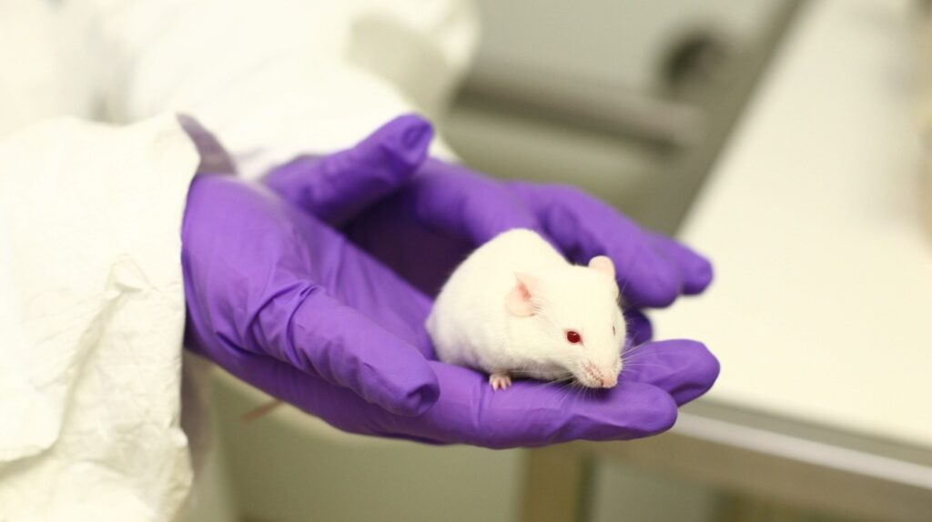
How animals are helping to improve our understanding of MND

Professor Lorenz Studer, who is the Director at the Center for Stem Cell Biology, Memorial Sloan Kettering Cancer Center in the USA, and one of our plenary speakers, will give an update on the technologies used in his lab to address some of the key challenges faced in using iPSCs, in order to realise their full potential in modelling disease and personalised medicine. His talk is titled ‘iPSCs as a model for neurodegenerative disease: Myths and truths (C15).
Dr Keiko Imamura will be talking about ‘ALS drug discovery using AI platform with patient iPSC panel’ (C16). During her research, Dr Imamura has used iSPCs to analyse the processes associated with the development of MND and screen therapeutic drugs using patient-derived motor neurons and an artificial intelligence (AI)-based drug discovery algorithm. This AI platform using patient iPSCs could be a powerful tool for discovering promising compounds for drug development, may make it possible to screen unlimited numbers of compounds.
Several projects funded by the MND Association have been studying different aspects of the disease using novel models. We look at two of them in closer detail.
Researchers from University College London have used a new mouse model to examine the characteristics and progression of ALS4, a slow progressing form of ALS with juvenile onset, caused by mutations in the gene that codes for the protein senataxin. To date, the mechanisms that underly ALS4 motor neuron degeneration are not well understood. This project aimed to improve this understanding (IVV-13).
Researchers from the University of Oxford have used iPSC-derived motor neurons and microglia (cells that help to maintain the health of the CNS) as a novel in vitro model of MND. There is growing evidence to suggest that inflammation plays a role in the development of MND, with microglia particularly implicated. However, their specific role – either harmful or protective – at different stages of the disease course and in different MND mutations remains unknown. Until now, microglial studies have been mostly limited to animal models and there remains the need for more authentic models using iPSCs. This project aimed to establish a novel in vitro model of inflammation in MND using both iPSC-derived microglia and motor neurons, and the findings suggest that this model is functional and will help to further our understanding of the role of microglia in MND (IVT-14).
Furthering our understanding of MND
Motor neurones are highly specialised, extremely long, single cells that require high levels of co-ordinated energy supply to perform their very complex role in the body. It is likely that their death in MND is caused by ‘attacks’ from multiple sources. Researchers are working hard to defend motor neurones ‘from all sides’ and, to do this, they need to know the neurones’ weaknesses.
Professor Julie Atkin is co-Director of the Macquarie Centre for MND Research in Australia. Proteostasis is the process that regulates proteins within the cell to maintain its health and involves a highly complex interconnection of pathways. Loss of proteostasis is implicated in the development of MND. In her talk ‘Dysfunction to proteostasis mechanisms in ALS/FTD’ (C9), Prof Atkins reviews recent studies linking intricate proteostasis mechanisms to MND and how therapeutic strategies based on restoring proteostasis may offer potential for future approaches for MND.

Unravelling and identifying the molecular events underlying motor neuron loss is of paramount importance if we are to increase the number of disease modifying therapies to treat MND. One such candidate is the protein WWOX, known to be involved in other neurological diseases. WWOX is involved in regulating the activity of some mitochondrial activities and in decreasing oxidative stress, implicated in the development of MND. A team of researchers from Boston in the USA set out to discover if alterations in WWOX contributes to mitochondrial dysfunction in MND. Their results suggest that loss of WWOX, as well as mutations in the gene, may cause and exacerbate mitochondrial dysfunction in MND, contributing to disease progression (IVT-19).
RELATED TOPIC
Blog | 16 March 2021 | Research Dev Team
Flexible molecules and droplets: researching and targeting RNA-protein complexes in MND
Researchers from the University of Bath, in this MND Association funded project, set out to understand the cellular and molecular mechanisms by which variants in the CFAP410 gene, identified as a risk gene for MND, contribute to the development of the disease, it at all. CFAP410 is a protein found at the base of primary cilia (microscopic sensory ‘antennae’ that cells use to gather information about their environment) and has been shown to interact with NEK, which is also found to be mutated in MND. Proteins associated with the basal body of primary cilia are increasingly being implicated in the DNA damage response, and the researchers found that this is the case with CFAP410. Mutations in the gene do not affect formation of the primary cilia but cause death of motor neurons by affecting the DNA damage response (IVV-29).
RELATED TOPIC
Blog | 21 April 2021 | Research Dev Team
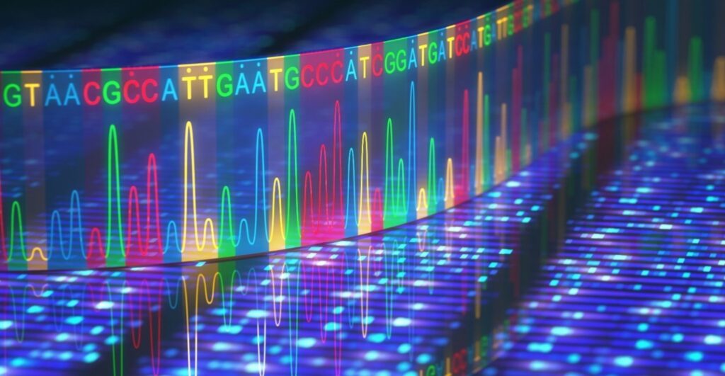
New insights into MND: 5 more genetic variants found
Stay informed
If you have come across this blog through the Symposium website, or a general search please subscribe (see top right hand corner of page) and you’ll be notified every time we upload a new article.
You can follow our research account on Twitter. We tweet about up to the minute research and will be tweeting throughout the Symposium – #alsmndsymp #drivingmndresearch
Take a look at the schedule of blogs for November as we continue counting down to the 32nd International Symposium on ALS/MND with our ‘Symposium Blogathon’.
To listen to talks live, take part in the Q&As and visit the live poster sessions, register for the International Symposium now.