My name is Tatyana Shelkovnikova, I am an MND Association Lady Edith Wolfson Senior Non-Clinical fellow. I lead my own group at the Medicines Discovery Institute, Cardiff University. I chose to move my fellowship to a drug discovery unit with a strong neuroscience portfolio of projects to be able to stay at the frontier of therapeutic innovation in this area. Although we have a fairly good view of MND causes, we now need to refine this knowledge and identify therapeutically promising molecules. I am still learning how drug discovery process works but I hope that in several years’ time, I’ll be able to use my research outcomes for patient benefit. I believe that MND is one of the most challenging of neurodegenerative diseases to tackle as a researcher. That is why every step forward by the global MND research community matters and makes us closer to a therapy.
Patients, donors and the Association team motivate me and keep my research going. I really appreciate the Association for continuing support in so many ways including financial backing for my research; it means so much to me and my team.
About my journey
I developed an interest in biosciences very early. Since the age of 12, I was taking part in biology competitions, having won prizes at a number of country-level ones. It predetermined my career choice, and I never imagined myself as anything other than a scientist. I have been involved in biomedical research since 2008. It was during my PhD studies when the genetic discovery of the faulty metabolism of ribonucleic acid (RNA) as one of the causes of MND was made, and this mechanism immediately attracted my attention. It was almost as if this specific field of study found me, and it has been at the core of my work since then. As a postdoctoral scientist, I was supported by a junior fellowship to get insight into the connection between intracellular droplets made of RNA and protein called paraspeckles and motor neuron malfunction. In 2018, I received funding from the Association to develop these findings and hopefully turn them into a therapeutic benefit, with the help of teams at the Cardiff’s Medicines Discovery Institute.
Last year, I was selected as the winner of the European Network for the Cure of ALS (ENCALS) Young Investigator Award 2020, adding to the list of MND Association-funded Research Fellows to win this prestigious award in recent years. I am delighted that my efforts to improve our knowledge of MND have been noticed and appreciated. Equally, I am humbled because I am very well aware of how many brilliant researchers and clinicians work hard towards the same cause.
Our work
We and others found that RNA-protein droplets found the cell nucleus – paraspeckles – can play an important role in protecting motor neurons from stresses and ultimately helping them survive.
Paraspeckles are prototypic “biomolecular condensates” – membrane-free structures inside cells that are formed by separation of liquid phases, just like oil separates from water. At the same time, paraspeckles are not mere droplets but instead have a very ordered internal structure. The main building block for paraspeckles is the RNA called NEAT1. Unlike many other RNAs, NEAT1 does not make a protein and its only known function is to assemble paraspeckles.
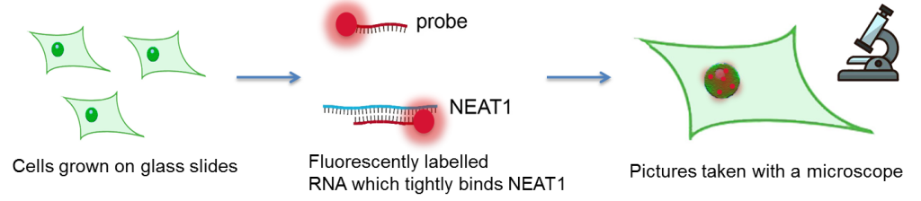
We can easily detect paraspeckles in cells, including neurons, by using RNA molecules called the “probe” – they bind to NEAT1 and bear light-producing beacons detectable by microscope. NEAT1 is a proxy for paraspeckles.
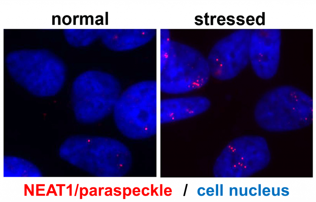
Paraspeckles were discovered about 20 years ago, but their normal function still remains largely unknown. What we do know is that they regulate the function of our genes, but which types of genes and how – is yet to be understood. Available data suggest that paraspeckles coordinate the action of multiple genes simultaneously. When cells are stressed, paraspeckle numbers increase dramatically, and when stress is resolved, paraspeckles almost disappear.
We and others found that survived motor neurons in the spinal cord of MND patients contain a lot more paraspeckles than neurons in healthy individuals. This prompted us to hypothesise that enhanced paraspeckles change function of neurons in a way that makes them more resilient and stress-resistant. However, this is still a hypothesis, and we use various models – human neurons growing in a dish and mice with altered NEAT1 levels – to test this hypothesis. Whilst it is clear that extra paraspeckles in neurons are there for a reason, what they do there is yet to be determined. Our recent finding is that paraspeckle accumulation is very prominent in chronically stressed motor neurons, where neurons in a dish endure mild stress for up to 72 hours. This may suggest that paraspeckles are the “last resort” for motor neurons which is activated when other protective resources have been exhausted.
Most importantly, increased paraspeckle formation is typical for MND cases regardless of disease cause – it is typical for familial and sporadic cases alike. This makes us believe that paraspeckle formation is a “disease signature” which can be used as a therapeutic target across different MND subtypes. With the disease being so heterogeneous, it is of paramount importance to identify such “universal” targets.
Next steps
Boosting paraspeckles in a targeted way, without compromising other processes in neurons, may help achieve a neuroprotective effect, and this therapy would be suitable for the vast majority of MND cases. One way of doing it is to find small molecule drugs that bind to paraspeckles and make them more stable. Since NEAT1 is the backbone for paraspeckles and is not found in any other structure in cells, it is a perfect molecular target.
Small molecule therapeutics have been traditionally viewed as less suitable for RNA targets since the latter are very dynamic molecules which are not expected to be tightly and specifically bound by small molecules. However, modern drug discovery science has found new avenues to increase the chance of RNA binding by small molecules. With NEAT1, we are fortunate because it contains a structure which is very much suitable for drug binding.
We have developed an approach to conduct rapid analysis (‘screening’) of chemical compounds in a test plate which would inform us on NEAT1 binding. Using this novel approach, we are currently scrutinising a library of thousands of chemicals to find NEAT1 binders. Once they are found, we will be able to test them in cells and make sure that they modulate paraspeckles in a meaningful way. These molecules (known as “tool molecules”) will be initially used in cells, including patient cells, to further study the formation and function of paraspeckles, before being modified and improved by medicinal chemists to make them suitable for human use.
We use the International Symposium on ALS/MND, organised by the MND Association, as the main forum to disseminate our findings and find new collaborations. Postdoctoral researcher from the lab, Dr Haiyan An, was a speaker at the 2019 Symposium in Perth, and presented our research on paraspeckles as a poster at last’s year’s virtual event. Her experience with the virtual presentation was very positive, with a lot of interest and engagement from early career researchers, such as PhD students.
Of course, we are still at the beginning of the drug discovery journey for NEAT1 and paraspeckles, which can span many years before translating into patient impact. But we are hopeful that these efforts will pay off and eventually make a difference for those living with this devastating disease.
We’d like to thank Tatyana for sharing her work in this blog piece, and congratulations for the well-deserved success of the ENCALS Young Investigator Award 2020.
We would also like to thank the Wolfson Foundation for their generous support of this fellowship.
Resources:
- ENCALS website: Young Investigator Award 2020
- MND Association website: Our Fellows
- MND Association website: Research we fund
- Cardiff University website: About us – Medicines Discovery Institute
- MND Research blog: How animals are helping to improve our understanding of MND