When diagnosing MND, it is important to look at the activity and impact of the motor neurones themselves – is the electrical message being carried down the nerve properly, and is it reaching the end of the nerve in the muscle? Malfunctions in the electrical activity at the muscle end of the nerve cell result in the muscle twitching that many people with MND experience.
One of the tests used to diagnose MND is an electromyography or EMG test. It involves putting needles into a muscle to measure electrical activity. It can be a painful and unpleasant experience, which doctors and patients are only willing to do when necessary.
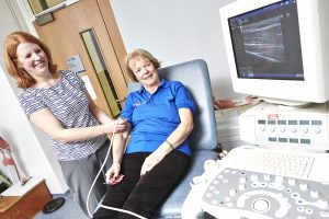
There is evidence that ultrasound imaging may be able to detect the same malfunctions in the electrical activity of muscle as EMG, by looking at the way the muscle behaves when electrical activity occurs. Ultrasound images produce the typical grey scale images, for example pictures from baby scans, and can be used to provide images of any muscles in the body.
Can ultrasound by used as an alternative to EMG?
Manchester Metropolitan University based Dr Emma Hodson-Tole and a PhD student in her lab Kate Bibbings have been investigating whether computational analysis of ultrasound images could provide measurements of muscle behaviour which would complement, or possibly replace EMG.
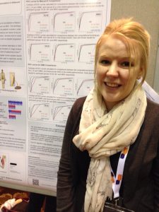
This research is funded by the MND Association ( our reference 866-792) and involves a team of researchers from MMU (Prof. Ian Loram, Dr. Pete Harding) and Dr. Nicholas Combes (Consultant Neurophysiologist, Royal Preston Hospital). Kate’s research is going well and she has presented her results to colleagues at conferences, and also to the nearby local branches of the MND Association.
There are several strands to Kate’s work, these include: ensuring that the ultrasound readings are consistently collected, that the readings are analysed and interpreted in a useful way and of course, importantly that ultrasound can distinguish between activity in healthy muscles as opposed to muscles affected by MND.
Making measurements more reproducible
If ultrasound is going to be routinely used, an automated computer based method of measuring the signals will be important. This will ensure that a wider range of people can conduct the tests, and it will reduce any differences in the way the results are interpreted. Developing and testing ways to do this has been a key part of Kate’s PhD. At the beginning of the project there was only one computer based approach for automatically measuring and analysing the ultrasound information. Kate has developed and tested two more.
Early work compared the results of computer based and manual identification of muscle activity in collected ultrasound images and found that the results match up well. Kate has also compared the results of muscle activity using ultrasound to the activity measured by EMG. Again the results compare well. All of the analyses have been conducted on willing volunteers, including people with MND.
Kate commented that: “It is always a pleasure to work with external participants when conducting our research and it had been clear from the start how enthusiastic and interested in the science behind the study our participants have been. We have also never had a shortage of people willing to get involved; even though the development of this technique is unlikely to benefit themselves, due to it being a diagnostic method.”
In the last year of her project Kate is working on ensuring the fine detail of the ultrasound technique is worked out – being able to identify the type of muscle movement, the level or amount of muscle movement and how it is different in different muscle groups.
Throughout June 2016 MND Awareness Month will be highlighting the rapid progression of the disease in its powerful Shortened Stories campaign, sharing the experiences of people currently living with MND, or who have lost loved ones to the disease, through art, poetry and film.