My name is Stephanie Shepheard, and I am a Research Associate in the Motor Neuron Disease & Neurotrophic Research Laboratory (MND&NR Lab), Flinders Health & Medicine Research Institute in the College of Medicine & Public Health at Flinders University, South Australia. I measure biological changes, or biomarkers, that may tell us something useful about motor neuron disease (MND) and translate these from the laboratory to practical tests in drug development clinical trials.
Diagnosing and monitoring MND is based on a person’s physical signs and symptoms, but MND is known for how widely these can vary between individuals. This makes it difficult to predict how quickly MND may progress, and what treatments may be useful, for any one person. It is difficult to design clinical trials that can detect a treatment benefit in lots of different people for the same reason. Thus, monitoring substances in body fluids and grouping people with the most similar biochemical signals, can assist in more easily detecting a beneficial treatment for MND. These types of signals are called biomarkers.
Biomarkers can be classified into different types based on what information they provide. This includes prognostic markers, which can help predict future changes in disease. A prognostic biomarker could ensure that people with a similar disease course are grouped together in clinical trials, making trial results clearer. For example, some SOD1 gene mutations are linked to quicker disease with a shorter survival.
Pharmacodynamic biomarkers are those that change in response to a potential treatment. For example, SOD1 protein in spinal fluid decreased when participants were given treatment in the Tofersen trial1. A subset of pharmacodynamic markers may also be biomarkers of disease progression, markers that change as disease progresses and in response to treatments. Thus, in clinical trials prognostic biomarkers would be used to group similar patients, and pharmacodynamic biomarkers to detect a benefit from treatment.
RELATED TOPIC
Blog | 21 July 2021 | Eleanor Green
Updates on Tofersen trials for SOD1-MND
My journey
My first laboratory experience was collecting and testing mouse urine! Specifically, the urine from a mouse model of MND in A/Prof Mary-Louise Rogers’ MND&NR Lab. Research by other groups had found two things of interest, first, that a protein called p75 is made by motor neurons (nerve cells that control movement) when they are damaged; and second, that part of the p75 protein is found in urine of rats who suffered spinal injury. Putting this together, we hypothesised that urine of SOD1G93A MND mice had high levels of p75ECD, and indeed, we were the first to show that p75ECD was higher in the urine of sick MND mice than their healthy counterparts, suggesting p75ECD could be a useful biomarker for MND2.
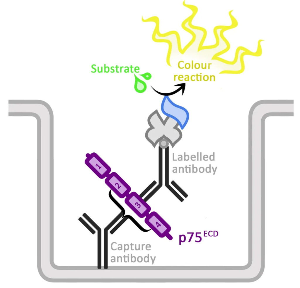
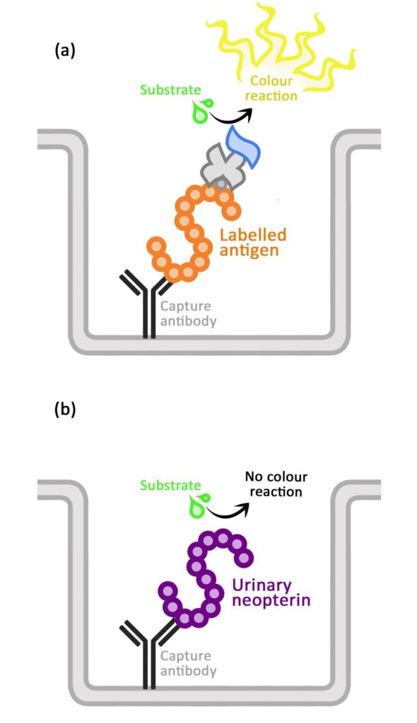
In my PhD I was the first to develop a test to measure human and mouse urine p75ECD using a technique called a sandwich ELISA (Figure 1A). With this I measured urine of people with MND and compared it to healthy people and people with other neurological conditions. Like the mouse model, we found that p75ECD was higher in people with MND, and interestingly, when looking at clinical information and p75ECD together, p75ECD increased in individuals as their disease symptoms progressed3. This means that urine p75ECD levels are a biomarker of disease progression in MND and may also be useful as a pharmacodynamic marker.
While p75ECD research continued in Australia, I joined the MND Association-funded AMBRoSIA (A Multicentre Biomarker Resource Strategy In ALS) study at the Sheffield Institute of Translational Neuroscience (SITraN) with Prof Dame Pamela Shaw. The aim of AMBRoSIA is to create a bank of blood, skin, urine, and spinal fluid samples from people with MND throughout their disease through collaboration between researchers and clinicians from Sheffield, Oxford (Prof Martin Turner and team) and London (Prof Andrea Malaspina and team). This bank of samples is allowing us to test potential MND biomarkers using many different techniques including p75ECD, and how often MND linked gene changes occur4.
Our team
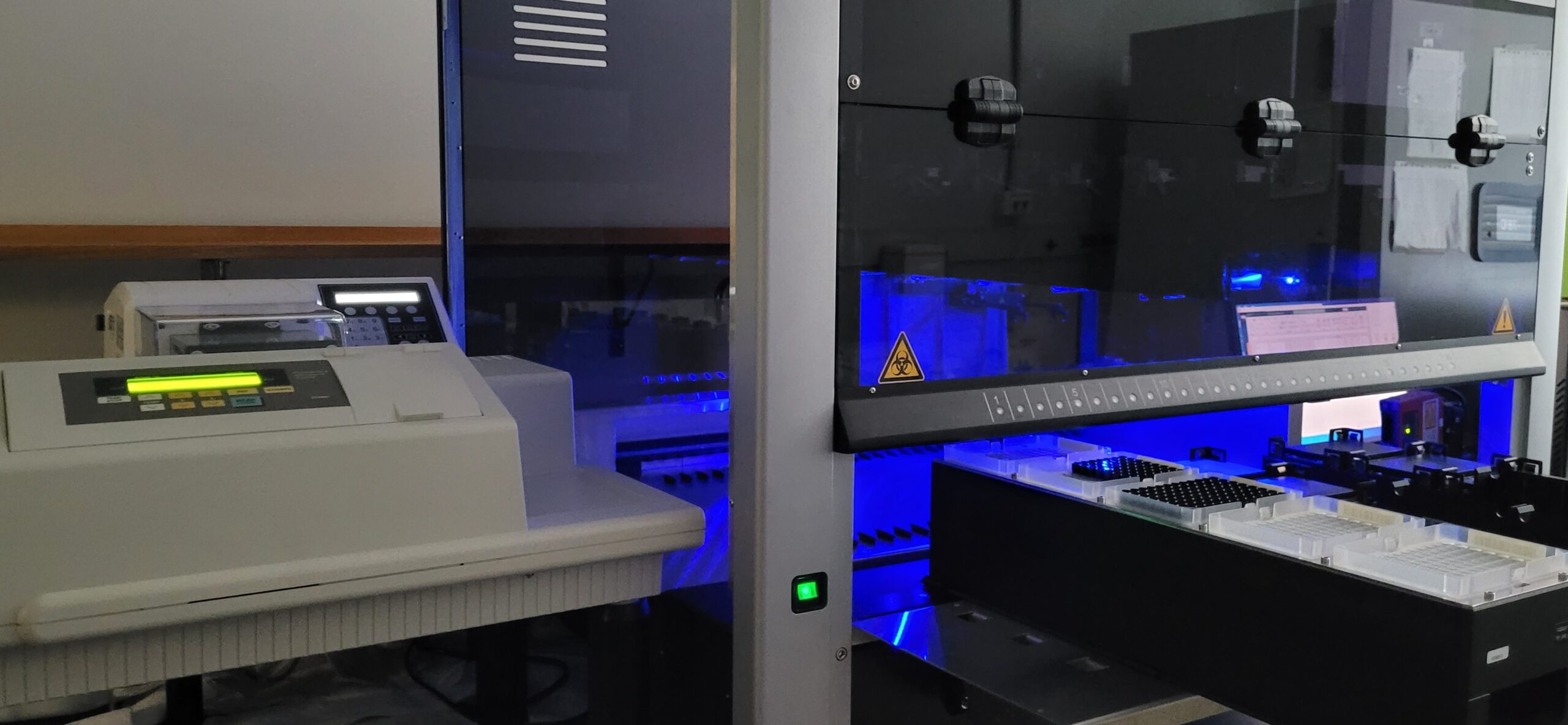
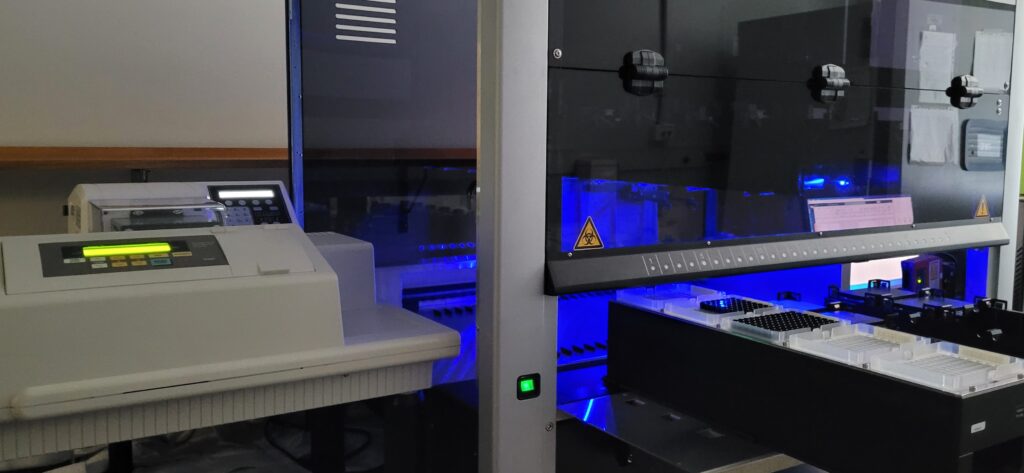
I then returned to Flinders University, where the ELISA has been upgraded to a specific human p75ECD ELISA robot setup (Figure 2), and I run the testing of urine samples from clinical trials and research collaborations (such as the CrEATe consortium & Prof Benatar) with A/Prof Rogers. Testing p75ECD in clinical trials is very exciting, as we can determine if it is useful as a pharmacodynamic marker. The robot also enables us to test samples much quicker, allowing our team more time to build a larger panel of biomarkers which tell us about different parts of disease.
While p75ECD is a biomarker of damage to motor neurons and other cells, many other processes in the body go awry in MND. One of these is the immune system, where it is hypothesised that the protective anti-inflammation environment becomes inefficient as motor neurons and other cells become damaged. The immune response then changes to a damaging pro-inflammation process which contributes to worsening disease. Several markers of the pro-inflammatory response have been researched by others in spinal fluid and blood, but not many in urine.
In our recent study we tested a small molecule called neopterin as a pro-inflammation biomarker. We know from research by others that neopterin is released by the immune system in the damaging pro-inflammation state and found in urine in the clinic in this state (including in COVID-19). This time, we used an inhibition ELISA rather than the sandwich ELISA we use for p75ECD (Figure 1B).
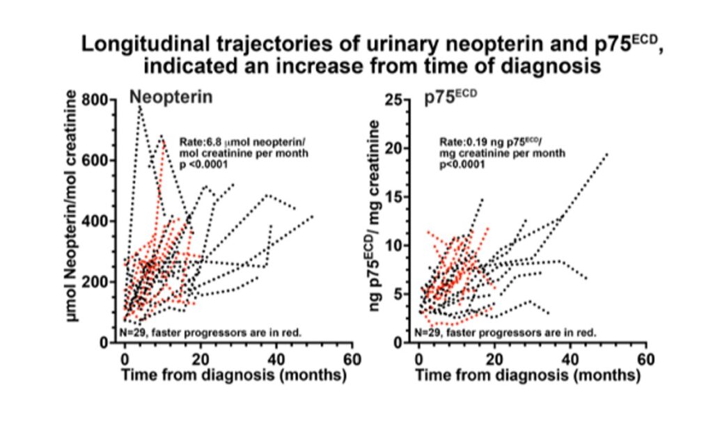
Testing urinary neopterin, and clinical information from participants in Australia and America along with p75ECD we found that neopterin is increased in the urine of people with MND and increases as the disease progresses, suggesting neopterin may also be useful as a disease progression biomarker along with p75ECD (Figure 3)5.
Our next steps with neopterin are the subject of Vassilios Karnaros’ PhD, in which larger groups of people are being tested and a high-performance liquid chromatography mass spectrometry method is being developed for neopterin testing. We then hope to use this method to quickly measure neopterin in clinical trials of drugs that target inflammation to see if it is a pharmacodynamic marker.
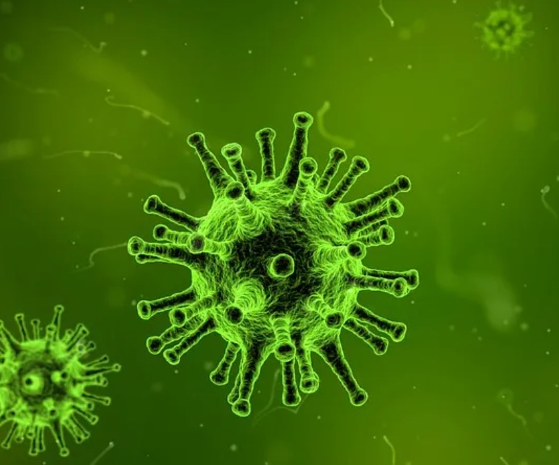
Another arm of our research program is the impact of human endogenous retroviruses (HERVs) in MND. It is hypothesised that MND may be caused or triggered by HERV-K, one of the types of viruses which infected animals and humans over millions of years of evolution and have since become part of our genes. Other researchers have found that genetic components coded by HERV-K are higher than normal in brain samples from people with MND, and that activating HERV-K genes killed healthy human neurons in experimental conditions. The Lighthouse clinical trials are investigating anti-retroviral treatment for MND, and PhD student Megan Dubowsky is determining if this anti-retroviral impacts the TDP-43 protein changes and immune dysfunction known to occur in MND using a mouse model. So far, we have found that MND mice treated with the anti-retrovirals have improved movement compared to the untreated mice, and we are continuing to research the immune and TDP-43 involvement.
RELATED TOPIC
Blog | 17 July 2019 | Martina Slapkova
Could MND be treated by HIV drugs?
Our research program is unique in using the non-invasive biomarker source of urine for potential MND biomarkers in models and people with MND. We are hopeful that our basic research and work in clinical trials will provide useful in drug development and clinical trials in MND.
References
- Miller et al., 2020, NEJM
- Shepheard et al., 2014, PLoS One
- Shepheard et al., 2017, Neurology
- Shepheard, et al., 2020, J Neurol Neurosurg Psychiatry
- Shepheard & Karnaros et al., 2022, Europ J Neurology