

Researchers from the University of Aberdeen and the University of Edinburgh have developed a new tool that could help to detect signs of MND in cells before symptoms of the disease start. This work was recently published and was previously presented at the 34th International Symposium in Basel by Dr Jenna Gregory.
The new tool is designed to identify and measure levels of a protein, called TDP-43, in tissue and biological fluids. TDP-43 is a known to become faulty in around 97% of people with MND and forms toxic clumps within cells in the body, including in motor neurons. In this study, the researchers tested their new tool, called an aptamer, to see whether it could help to uncover early signs of the disease and detect faulty TDP-43 earlier and with more sensitivity than current methods.
What is an aptamer?
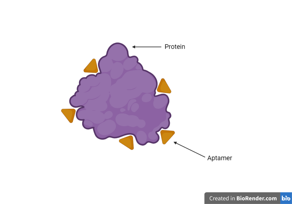
An aptamer is made from the same material that our DNA is made from, called nucleic acid. It is a chemical that is designed and made by researchers to attach to a certain protein or structure within a cell. This approach allows the protein or structure to be identified or measured more accurately and easily using a microscope. Researchers can use aptamers to visualise and measure proteins and structures within tissue samples. They bind to more than one site on the protein or structure that they are designed to detect. Aptamers have been used in the treatment and diagnosis of other diseases for several years. For example, they are used in cancer diagnosis to identify proteins and other compounds which are made by cancer cells. However, they aren’t currently used in MND.
What are the current methods for detecting TDP-43?
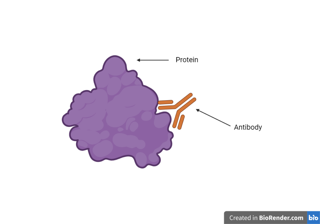
Current methods for detecting TDP-43 in tissue relies upon the use of antibodies. Antibodies are proteins which can recognise and bind to particular parts of other proteins. For example, antibodies to detect the TDP-43 protein can be used to visualise where TDP-43 is in the cell, show whether it has formed clumps and help to measure levels of it. Antibodies usually bind to one specific site on the protein or structure that they are being used to detect.
What did this work focus on?
This study focused on using the newly developed aptamer to observe faulty TDP-43 in MND and test whether it could be a good alternative to antibodies.
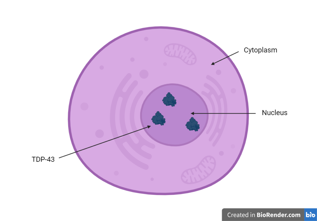
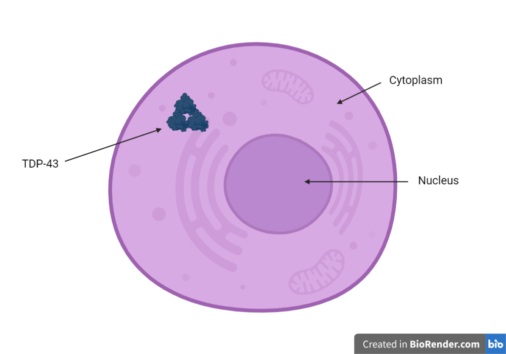
They also wanted to understand more about the role that faulty TDP-43 plays in MND. In MND, TDP-43 becomes faulty, moves out of the nucleus of the cell (control centre of the cell), where it needs to be to work properly, into the cytoplasm (main body) of the cell. It forms clumps within the cytoplasm which are toxic and contribute to cell damage and death.
In this study, the researchers wanted to use the new aptamer to observe how faulty TDP-43 behaves in both locations in the cells and uncover more about how it is involved in MND. To do this, they used postmortem brain tissue from 24 people who had MND and healthy controls from the Edinburgh brain bank. The researchers added the new aptamer and antibodies to the tissue samples to compare the methods and observe TDP-43 in the cells.
Why was this work needed?
Current antibody detection methods may not be sensitive enough to show early signs of TDP-43 pathology. This is because in MND the faulty TDP-43 protein forms clumps and the area that the antibody is designed to bind to may be buried inside the clump. This means that the antibody may no longer be able to bind to TDP-43 and researchers might not be able to accurately observe or measure levels of the faulty protein. This study tested the new aptamer method to see if it could help to detect early signs of faulty TDP-43 in MND and allow researchers to observe levels of the protein more accurately in tissue samples.
What did the researchers find?
In this study, the researchers found that the aptamer could successfully detect faulty TDP-43 in the samples of brain tissue. It also did not detect any faulty protein in the tissue samples from the healthy controls. This suggests that the aptamer is specific as it only binds to TDP-43 when it is faulty and helps to highlight where it is located in the cells. Due to its ability to only detect faulty TDP-43, it may be useful in diagnosing MND in the future.
The researchers demonstrated that the aptamer can be used to observe much smaller clumps of faulty TDP-43 than the current antibody method. The presence of smaller clumps of TDP-43 are an early sign of cell damage and using the aptamer to detect these in tissue might mean that MND can be diagnosed earlier in the disease course. This shows that the aptamer is a lot more sensitive at detecting faulty TDP-43 than the antibodies.
As well as testing the new tool, the researchers wanted to use it to try and understand more about the behaviour of faulty TDP-43 in MND. They discovered that faulty TDP-43 also occurs in the nucleus of the cells before it moves to the cytoplasm. It is thought that faulty TDP-43 in the nucleus may be a very early sign of disease and might even be present before symptoms of the disease begin. The study also found that clumps of TDP-43 in the cytoplasm are likely to form after symptoms of MND have begun. This has helped to shed more light on the role that faulty TDP-43 may play in the development of MND and highlight the importance of understanding more about faulty TDP-43 in the nucleus.
What does this mean?
MND remains difficult to diagnose due to the initial symptoms of the disease being similar to those seen in other neurological conditions. As there is no specific test for MND, other conditions that may cause these symptoms have to be ruled out before the clinician can be sure someone has MND. This means that, on average, people currently receive a diagnosis a year after the initial onset of symptoms. The new aptamer tool could be used alongside other tests to help to diagnose MND earlier in people who are experiencing symptoms of the disease. Earlier diagnoses could mean that more people with MND are eligible to take part in clinical trials and can access treatments, care and support sooner.
The study has also helped to increase current understanding of the role that faulty TDP-43 may play in the development and onset of the disease. It has suggested that it may be possible to use the aptamer to detect very early signs of MND, before symptoms occur, by looking for faulty TDP-43 in the nucleus of the cell. If faulty TDP-43 in the nucleus can be confirmed as an early sign of the disease, researchers may be able to study ways to prevent further damage from occurring and delay or stop the movement of faulty TDP-43 into the cytoplasm, which seems to be linked to the onset of symptoms.
What might be the next steps for this research?
As the aptamer has been confirmed as a good alternative to antibodies for detecting faulty TDP-43, it may now be used in future research to help us to uncover more about the behaviour of TDP-43 in MND. This research highlighted that faulty TDP-43 can be seen in the nucleus before it clumps in the cytoplasm and that this may happen very early on in the disease before symptoms occur. However, further research is now needed to fully understand how faulty TDP-43 in the nucleus is involved in MND and exactly how the formation of the clumps in the cytoplasm are linked to the development of symptoms. If researchers can uncover more about the faulty TDP-43 in the nucleus, interventions might be able to be developed to reduce or stop clumps from forming in the cytoplasm, which may delay or prevent symptoms of MND.
This study has indicated that the aptamer might be a useful diagnostic tool and allow MND to be diagnosed earlier than it is currently. However, further research into how this could be used in the clinic is needed before it is used routinely. It is likely that this aptamer will be used alongside other tests to confirm a diagnosis of MND rather than being used on its own. The aptamer would need further testing to determine whether it could detect faulty TDP-43 in biological fluids of people with MND, such as blood or cerebrospinal fluids. This study only looked at the aptamer in brain tissue which cannot be taken from people with MND as part of routine tests in the clinic.
The aptamer in this study is an exciting new tool which can be used in research to help us build our knowledge of the role that TDP-43 plays in the disease and allows researchers to observe and measure the faulty protein in the lab more accurately. While this new tool is a very promising advancement in MND research and definitely a step in the right direction, a significant amount of further research is needed before this can be used in the clinic for people with MND. We hope that this new tool continues to be successful in research and can be used to detect faulty TDP-43 in biological fluids so that it can move towards being used routinely in MND clinics to aid in earlier diagnoses.