
Weight loss is a problem for many people living with MND, with approximately 20% of people already having lost 10% of their weight at diagnosis. Some of this weight loss may be due to a person experiencing difficulties with chewing and swallowing because of increased muscle weakness. It could be due to a lack of appetite because of tiredness, anxiety or low mood. It can also be caused by hypermetabolism which is when the body uses more energy than is being consumed, even without exercise, and is seen in some people with MND. Studies have shown that weight loss is linked to shorter survival.
At the 34th International Symposium on ALS/MND in Basel, Switzerland one of the sessions looked at nutritional assessment and management, with talks covering feeding tube management, the decision-making process of feeding tube placement, how diet might help slow progression, alternative ways to measure nutritional state and how appetite is controlled in the brain. In this series of five blogs, we are going to look a little closer at each of these studies and their potential for helping people living with MND. This is the final blog in the series.
Jeryn Chang, a PhD student from the University of Queensland, Australia discussed how appetite is controlled in the brain in people with MND and what effect this might have on their weight loss over the course of their disease.
Central pathways of appetite control in MND: fMRI evidence of altered brain responses to visual food stimuli
As we all know, MND is an incredibly varied disease and this is largely due to the different phenotypes in MND. However, over the years, there has been increased understanding that there are other factors involved, such as energy balance, that contribute to disease progression and survival. Examples of these include:
- Nutritional status
- Loss of weight
- Loss of fat mass
- Circulating lipids (fats)
- Hypermetabolism
- Fatigue
- Loss of appetite
Loss of appetite is particularly important, and it occurs in up to 30% of people with MND. Loss of appetite has been shown to be associated with weight loss and loss of fat mass leading to faster disease progression and shorter survival. So, it’s clinically important to discover the biological mechanisms underlying this.
What did they do?
The first place to look is the hypothalamus, which is a small area in the brain and is the central regulator for appetite and whole-body metabolism. Over the years there have been studies conducted that have looked at the hypothalamus in MND and have shown that the volume of the hypothalamus is decreased and that this has a positive correlation with body mass index (BMI). This might suggest that lower hypothalamic volume could lead to loss of appetite and metabolic regulation which, in turn, leads to loss of fat mass and shorter survival.
This was looked at in the participants in this study and hypothalamic volume was observed. Those with lower BMIs and lower hypothalamic volume tended to lose weight over the course of their disease leading to shorter survival. This really demonstrates the complex role the hypothalamus plays in energy regulation and appetite and the need to look beyond the hypothalamus to other regions of the brain as well.
The Appetitive Brain Network
There have previously been many studies that have looked at what is termed as the ‘appetitive brain network’ (Figure 1) – different areas of the brain that respond to food cues – particularly in the areas of obesity and anorexia nervosa, and these studies used functional MRI (fMRI) to assess brain response to visual stimuli of food items and high calorie food items and saw responses in different areas of the brain as shown in Figure 1.
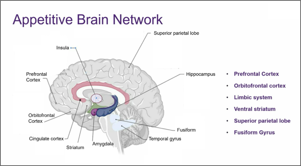
Figure 1: Illustration of the brain showing the Appetitive Brain Network. Reproduced from Dr Chang’s slides, International Symposium on ALS/MND 2023.
A similar study in MND hasn’t been conducted before. This study aimed to identify areas of the brain that respond to visual stimuli of food in people with MND compared to those without the condition, with the assumption that deficits in functional activity in known appetitive networks will be seen in the people with MND.
After selection, 35 people with MND moved forward to imaging. During the course of the study, anthropometric measures (weight, body composition), appetite measures and clinical measures (symptom onset and ALSFRS-R) were also collected. Data was also collected from 23 controls. Appetite was measured using the Council on Nutrition Appetite Questionnaire (CNAQ) and this showed that there was a significant decrease in appetite in people with MND compared to controls. However, the two groups were well matched in weight, BMI and percentage fat mass.
Participants were asked to fast the night before the scan and on the day. Participants then underwent two imaging sessions, the first under a fasting condition and the second after the consumption of a liquid mixed meal (a nutritionally complete meal substitute in liquid form). Within each imaging session, participants were shown randomised images of non-food items, low calorie foods and high calorie foods.
What did they find?
The first step was to see how participants reacted to non-food items. The images seen by fMRI showed activation in areas of the brain consistent with what is already known about visual processing. These images showed that there was no significant difference between the people with MND and the controls when shown images of non-food items, in both the fasting and non-fasting state, suggesting that both groups view pictures of non-food items very similarly.
When shown pictures of low and high calorie food items, regardless of whether this was before or after consumption of the liquid mixed meal, the images showed activation in the areas of the brain associated with the appetitive brain network. Again, there was no significant difference between each group.
Looking at the images of both groups in the just the fasted state, similar activation of the brain appetitive network was seen in all participants.
Attention was then given to images of high calorie food in the fasting state and no significant difference was observed between the groups.
This means that when both healthy individuals and those with MND look at pictures of foods their brain activity is similar, at least in the fasted state.
However, when shown pictures of high-calorie foods in the fasting state, the researchers noticed that in healthy individuals there’s increased brain activity in a specific area related to personal and emotional memories, called the right temporal pole. This increase is not seen in people with MND, whether they are hungry or full. This might suggest that the temporal pole is affected by eating food, but this response is impacted in people with MND. This difference in brain activity might be linked to the fact that people with MND often experience a decrease in appetite, as previously documented in the appetite questionnaires.
The researchers were also surprised to find that in hungry healthy individuals, appetite is associated with the cerebellum, a part of the brain known for motor control. But in people with MND, this connection between appetite and the cerebellum is weaker. This discovery was interesting because until recently, the temporal pole and cerebellum weren’t thought to be directly related to appetite and reward.
Recent evidence suggests that these brain areas might play a crucial role in regulating appetite and reward. Both the temporal pole and cerebellum are interconnected regions, especially within the limbic system, which is involved in emotions and rewards. Previous studies have also shown changes in these areas in individuals with MND, including altered connectivity in the temporal pole and increased functionality in the cerebellum during motor tasks, which might suggest the cerebellum plays a compensatory role in MND.
Going forward
The involvement of less well-known appetite regions has been observed in this study, which might suggest that alterations in appetite in MND may be more complex than previously thought. The researchers found it curious that deficits in activations in the temporal pole and cerebellum were seen, and future studies in these regions are needed to provide further insights into the functions and consequence in MND.
The International Alliance of ALS/MND Associations held a webinar in February 2024 that discussed nutrition in ALS/MND – its importance, nutritional changes after diagnosis, considerations for bulbar onset ALS/MND and the myths and benefits of feeding tubes. You can watch the video below.
We hope you have enjoyed this series of blogs discussing the latest research in weight control and management in MND. The Symposium is an amazing platform for researchers to share their work in this and other equally important areas of MND research. It generates discussion and debate, and the foundations of many great discoveries have been laid here. Please check out other blog articles covering talks from the International Symposium on ALS/MND 2023.
None of this research would be possible without the people with MND who take part, and those who support them. We thank them for their trust and time.