This blog is part of the ‘Highlights from Glasgow’ collection of articles, where you can read about the content of some of the talks and posters presented at the 29th International Symposium on ALS/MND.
Around the globe, teams are working to find tissue biomarkers for MND and there some promising candidates coming through, some of which were explored at session 8C of the Symposium: Tissue Biomarkers.
We first heard from Rebekah Ahmed (C83), who presented her data on possible neuroendocrine and metabolic biomarkers. Ahmed and her team analysed several proteins related to these systems in the blood of 127 people with either MND or MND with FTD and compared them to controls. The Neuropeptide Y protein (NPY), which is related to eating habits and food intake, was found to be higher in people with MND and MND-FTD, and lower in people with FTD, shining the light on a possible biomarker. All patients also had an increase in leptin and insulin levels, highlighting the need to examine how these neuroendocrine changes affect underlying disease pathology.
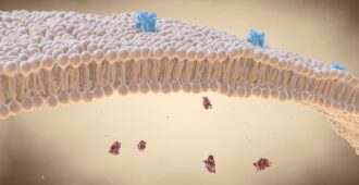
Next, Valentina Bonetto (C84) explored the possibility of using extracellular vesicles (EVs) as a biomarker of MND. EVs are like packages that can transport materials between cells, such as proteins and RNA, to facilitate waste disposal and communication between cells. They may also play a part in the spread of disease – several proteins linked to MND play a role in EV activity. An effective biomarker needs to be easy to measure, however, EVs are difficult to isolate and there is no gold standard yet on how to do this. Bonetto presented a new method that the team have developed to isolate plasma EVs, potentially overcoming this challenge. Analysis using this method revealed that people with MND had a significantly higher concentration of EVs compared to healthy people and people with other neurological diseases. The concentration was highest in those with the slow progressing form of MND, potentially showing a biomarker that can distinguish between fast and slow progressing forms of the disease. They also saw that people with MND had remarkably smaller EVs, and lower levels of a protein called PPIA, which warrants further investigation.
Pam Factor-Litvak (C85) looked at the levels of miRNAs that are packaged within EVs, which are heavily implicated in MND. Her study involved analysis of 100 samples from the United States National US Registry. She isolated miRNAs from the plasma of 100 patients and looked at their activity. Analysis showed that healthy controls, people with Multiple Sclerosis, and people with sporadic MND each had distinct inflammatory miRNA profiles (sets of miRNA that become more active). This suggests that analyzing these sets of miRNA could distinguish MND from other diseases.
Next, Greg Joilin (C86) presented on his findings of a unique signature of non-coding RNA from the serum of people with MND. He identified a set of non-coding RNA that could distinguish fast from slow progressing MND in 90% of the cases in the study. Joilin and the team are now carrying out further work to validate this promising signature.
Pierre-François Pradat (C87) next looked at ferroptosis – an emerging new cell death mechanism that occurs via a process of lipid peroxydation. Through studying over 100 patient samples from a recent clinical trial, his study supported that markers of ferroptosis are predictive of functional decline in MND (measured by the ALS-FRS).
Finally, Osamu Kano (C88) presented his findings which support that the neuronal apoptosis inhibitory protein (NAIP) is a risk factor and a prognostic biomarker in MND. Through analyzing the changes in NAIP levels over a period of 12 months, Kano found that people with a higher amount of NAIP had a smaller decline in function (measured by ALS-FRS) over 12 months, and vice versa. As NAIP is known to protect neuronal cells against oxidative stress, this suggests that high levels might be protective in MND as well as showing promise as a biomarker.
Further information on this topic was presented at the poster session (Theme 6) where data on many possible tissue biomarker signatures was presented, including a poster by Daisy Sproviero (BIO-18) and her team, who found a small group of RNAs in extracellular vesicles that were specific to MND.
Find out more about the topics discussed in Glasgow on our Periodic table of Symposium at www.mndassociation.org/symplive.


