Dr Gareth Wright, based at the University of Liverpool, is a postdoctoral researcher funded by the MND Association. His research is all about using physics and x-rays to further our understanding of MND. Here he gives us a taste of why X-rays are important.
The background to X-rays
We have a long history of X-ray science in Liverpool. In 1896 Sir Oliver Lodge used X-rays to image a lead pellet embedded in the hand of a 12 year old boy. This was one of the first medical uses of X-rays and allowed the bullet to be successfully removed. Charles Barkla made an observation in 1904 considered to be the birth of X-ray science; X-rays behave like visible light and are part of the electromagnetic spectrum. They have wavelength around 0.1 nm (0.000000001 metres!) which makes them perfect to resolve individual atoms in a molecule (eg water).

This year is the hundredth anniversary of the discovery that X-rays are diffracted by crystals. To mark the occasion, the United Nations declared 2014 the International Year of Crystallography. A huge amount of development has changed X-ray science in those hundred years. Our experiments now commonly take place at synchrotrons; large particle accelerators that speed electrons (negatively charged particles that are part of an atom) around a ring at close to the speed of light. This is an amazing feat in itself because the mass of an object increases as its speed increases. If a tennis ball were to travel at the same speed as our electron it would weigh roughly 4 Kg. The size and cost of these facilities means we can’t have one in every university, city or company. In the UK, Diamond in Oxfordshire is the national synchrotron. My group also regularly uses Soleil in France and SPring-8 in Japan.
Using X-rays to study proteins
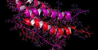
Synchrotron X-rays are produced when an electron’s trajectory (the path a moving object follows) is shifted in a magnetic field. The advantage of synchrotron X-rays is they are much brighter than those generated by traditional methods. We can use them to gather information about almost any substance we’re interested in. In my case that’s proteins which have a link to motor neurone disease. One technique I have used a lot in the last 5 years is small angle X-ray scattering (SAXS). Unlike crystallography where you need a crystal to collect data, SAXS can tell you a lot about the overall size and shape of a protein free in solution.
Interesting proteins are often difficult to work with. Some proteins stick to each other, some fall apart, others exist in different forms at the same time. Each poses a problem if you want to know something specific about one component. What’s special about my experiments is the combination of SAXS and chromatography. A chromatography stage allows me to separate everything in my sample immediately before exposure to X-rays. Many neurodegenerative disorders, particularly MND, can be caused by proteins that don’t form a single structure or that stick together in clumps. These proteins, such as SOD1, TDP-43 or FUS are perfect for this type of experiment.
From Liverpool to Paris
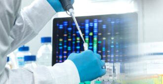
The work for a synchrotron trip begins many months in advance of the data collection. All my protein production and purification happens in Liverpool where I make sure the protein sample is in a good enough condition to get the information I need. Beam time for chromatographic SAXS is rarer than most as there are only a handful of stations worldwide where you can do these experiments. So I go to Paris with my samples. We usually have a 24 hour shift and 2 people will be there for safety. At 8am I arrive at the station and make all the solutions I need for the day then prepare the chromatographic system. A typical experiment begins by loading a protein sample onto the chromatographic system. As separation occurs and the protein is released it is fed in to the X-ray beam. Most X-rays go straight through the sample and carry on undisturbed but some interact with the protein, or the liquid, and change course very slightly. We record the amount and direction of these X-rays.
Understanding more about MND in just 24 hours
The data we get from a SAXS experiment is very simple but with some clever analysis it can tell you a lot about your protein. In the past I’ve used it to see structure changes, unfolding and aggregation of proteins involved in MND. All this helps us to understand why these proteins cause MND and gives us clues about what we can do to fix it.
Twenty-four hours sounds like a long shift but it goes pretty quick. Between preparing your sample, the agonising wait for your protein to hit the beam and data processing you don’t get a chance to think about how late or early it is. 8am comes around again and that’s it, time for a Parisian omelette or onion soup and back on the train to Liverpool.


Nice explanation!